

Advancing Retinal Diagnostics With Multimodal Imaging
Lena Labs develops advanced imaging technologies to improve the detection and analysis of retinal disease.
450 - 100
NM DETECTION RANGE
Visible to Near-IR
8
SPECTRAL CHANNELS
Simultaneous Capture
66 fps
REFLECTANCE MODE
Frame Rate
>97%
SPECTRAL ACCURACY
After AI Denoising
Multimodal Retinal Imaging
-
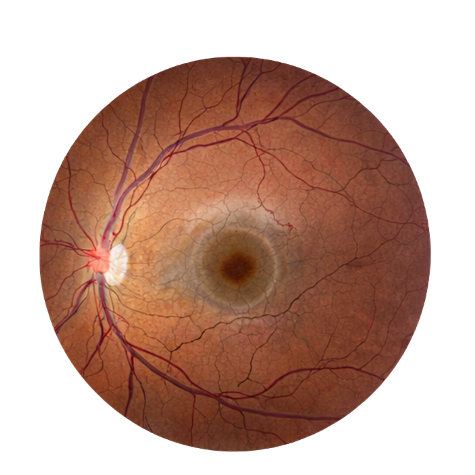
VISIBLE
.
-
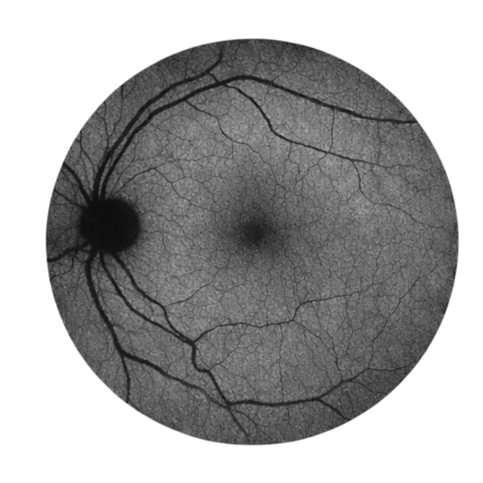
FAF
-
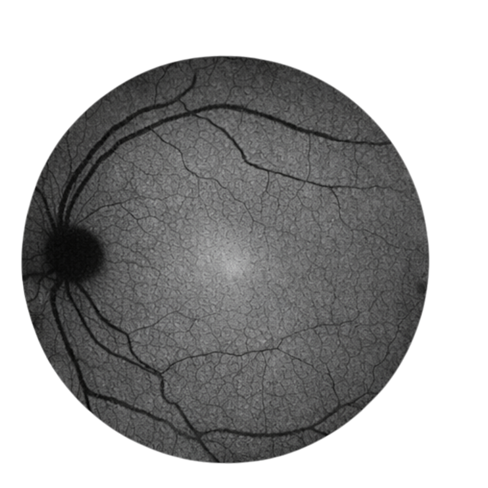
NIR
.
CORE TECHNOLOGY
Engineered for spectral precision
The DuOS (Dual-mode Optical Spectroscopy) system integrates spectral reflectance and autofluorescence imaging within a single optical pathway, combining optics, electronics, and AI to capture what current retinal cameras cannot see.
Multimodal Hyperspectral Imaging
Combines broadband reflectance and autofluorescence within a single optical pathway. Three discrete excitation bands — 488 nm, 560 nm, 780 nm — enable blue, green, and near-infrared autofluorescence without mechanical realignment.
AI-based spectral denoising algorithms recover clinically usable signals under low-light imaging conditions while preserving spectral fidelity. This approach enhances signal quality without compromising spectral accuracy.
Broad Spectral Snapshot Detection
The system captures a wide spectral range spanning from visible to near-infrared wavelengths (400–1000 nm) within a single snapshot acquisition. This extended spectral capability supports enhanced biochemical and structural analysis of retinal biomarkers.
Integrated Calibration & Real-Time Spectral Processing
Integrated calibration and processing software enable real-time spectral analysis within a unified platform. The system supports consistent spectral performance and efficient data processing across imaging sessions.
LENA LABS
Lena Labs focuses on the development of multimodal spectral imaging systems for retinal diagnostics and clinical research.
Its flagship platform, the Lena DuOS (Dual-mode Optical Spectroscopy) System, integrates spectral reflectance and autofluorescence imaging within a compact architecture to support comprehensive retinal tissue characterization.
Clinical Applications
Revealing what conventional imaging cannot

Age-Related Macular Degeneration
Spectral reflectance and autofluorescence signatures enable quantification of drusen, lipofuscin, and RPE integrity — biomarkers that remain invisible to standard color fundus photography.
Geographic Atrophy
Spectral reflectance and autofluorescence signatures enable quantification of drusen, lipofuscin, and RPE integrity — biomarkers that remain invisible to standard color fundus photography.